Interesting Radiographs
Learn a little more on how and why we use radiographs below.
Radiology 101
Radiographs work just like a film camera. X-rays are passed through the patient to expose a piece of radiographic film on the other side of the patient. Thin areas of the patient allow a lot of X-rays through, which exposes the film and turns it black. Thicker body parts absorb more X-rays, so less hits the film, resulting in white areas. Thus white objects are dense like bone, and black objects are thin like air. It is normal for animals to swallow air when eating, so there is always a little air in the stomach or intestinal tract. This shows up as black ribbons of various sizes in the abdomen.
How Many Puppies?
This dog’s owner knew she was pregnant and was hoping for a big litter. Can you count how many puppies she is expecting? Here’s a hint: the little puppy skeletons are visible. Try to count heads and backbones. It’s harder than it looks because the puppies overlap each other!
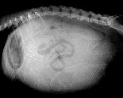
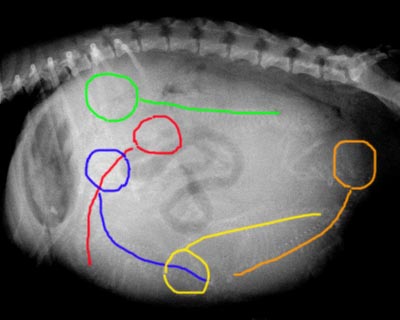
HERE IS THE ANSWER:
There are five puppies. We have outlined the heads and backbones in different colors to point them out. Compare this image with the uncolored one above. How did you do?
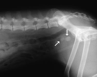
BLADDER STONES
Bladder stones (also known as uroliths or cystic calculi) are hard, rock-like lumps of mineral that form in the urinary tract. People usually get stones in the kidneys that are small and can be passed, although the process is painful. Dogs and cats are different and usually form stones in the bladder. Because it is a bigger space, these stones tend to be much larger and usually cannot be passed but have to be removed surgically or dissolved with special diets.
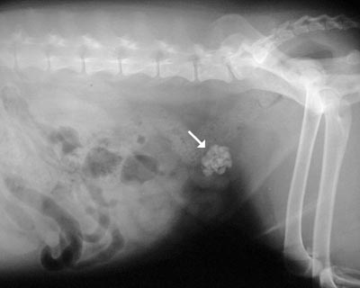
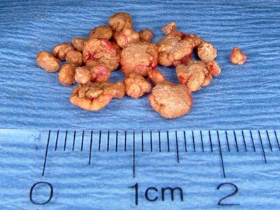
We suspect bladder stones when a dog has blood in the urine that doesn’t respond to conventional antibiotic therapy, when we palpate a thickened bladder, when a dog strains to urinate, or when there are many crystals in a urine sample. Because most stones contain calcium, they are usually visible on a radiograph as white (bone density) objects in the area of the bladder. Sometimes there is a single large stone, and sometimes there are literally hundreds of tiny stones like sand or aquarium gravel. The radiographs show typical cases of bladder stones. The second photo shows some stones after surgical removal from a dog’s bladder.
When Animals Limp
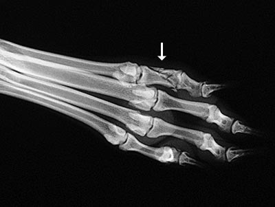
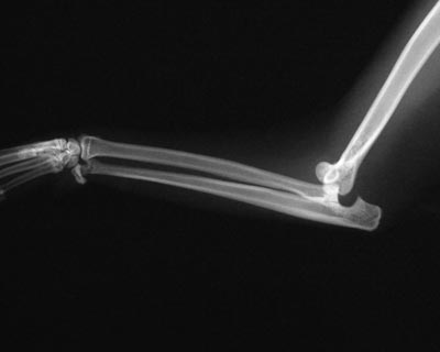
The good news is that 9 out of 10 limping animals have what we call soft tissue injuries (i.e., a sprain, a pull, a bruise) and only require exercise restriction to heal. However, 1 out of 10 animals with lameness has something more serious, and we can tell which ones by close observation of the patient walking and careful physical examination. In those cases, radiographs are used to make a diagnosis. The dog featured in the photo had a badly broken toe and was treated with a splint. The cat featured in the other photo fell out of a tree and dislocated its elbow. The injury was so severe that surgery was needed to put it back together.
HIP DYSPLASIA
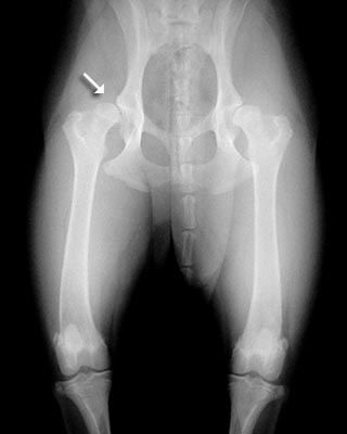
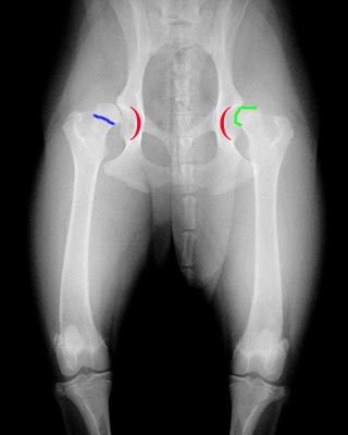
Hip dysplasia is a looseness in the hip joint. The hip is a ball-and-socket joint, and the head (ball) of the femur (thigh bone) normally should be deep within the hip socket. When hip dysplasia is present, the ball moves in and out of the socket with ease. Over time arthritis (degenerative joint disease, osteoarthritis) sets in as the body tries to stabilize the loose joint. You can see both hips are loose in the photo, although the left one is worse than the right. The sockets are outlined in red to show where the head of the femur should be. The head should be perfectly round but often becomes flattened and angular as arthritis develops. The green line outlines this; check out that area on the uncolored radiograph as well. A white line across the neck of the femur (the narrow part between the head and the shaft of the femur) outlined in blue is another early sign of arthritis. Dysplasia is primarily an inherited condition but can be made worse by certain environmental factors during growth.
WHEN LAMENESS ISN’T SIMPLE
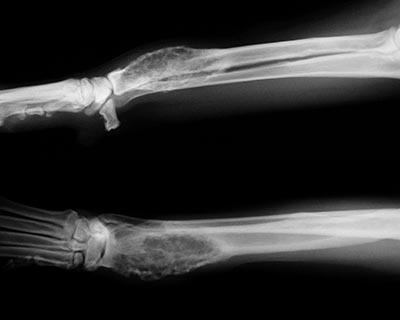
Sometimes when an animal limps the cause turns out to be something more serious than a simple injury. The doctors palpated a firm, painful lump in the leg this dog was favoring. Radiographs showed that the bone was expanded in that area, with a moth-eaten, hollowed-out center. These are classic signs of a tumor in the bone, known as an osteosarcoma.

